


Description
For Biomolecule Detection


Nitrocellulose membrane is commonly used for nucleic acid and protein detection in research and diagnostic applications. Nitrocellulose membrane from Pall is a pure 100% unsupported nitrocellulose media that has high binding capacity for nucleic acids and proteins. The high protein binding capacity is ensured due to the homogenous nature of the membrane. Other nitrocellulose membranes contain high levels of cellulose acetate, which reduce the protein binding capacity of the membrane. Physical characteristics of the nitrocellulose membrane from Pall, such as high tensile strength and hydrophilicity, provide excellent handling ensuring that the membrane will not rip, tear, or crack during transfers. The detergent free and unsupported nature of this material yields sharper images with little or no distortion. Compatible with a variety of detection systems, it has lower protein burn through than competitive nitrocellulose media in electrophoretic transfers. Highly consistent, both inter- and intra-lot, this membrane will provide sensitive detection of biomolecules blot after blot.
Specifications
Typical Membrane Characteristics
| Base Material | Pore Size (µm) | Thickness (mils) | Thickness (µm) |
| Nitrocellulose (BioTrace& NT) | 0.2 | 4.0-7.5 | 101.6-190.5 |
Typical Performance Characteristics
| Base Material | Binding Interaction | Immobilization Methods | Detection Methods | Wet Time (sec) |
| Nitrocellulose (BioTrace& NT) | Hydrophobic, electrostatic | Electrophoretic transfer, direct spotting, capillary transfer, dot blotting | Chemiluminescence, fluorescence, direct staining, radiolabeling, colorimetric | < 5.0 |
*Biosafety: complies with United States Pharmacopeia (USP) Biological Reactivity Test, In Vivo <88> for biosafety, cytotoxicity, and hemolysis testing.
The BioTrace name is a trademark of Pall Corporation and is not available for use.
Applications
Applications
- Western blots
- Southern blots
- Northern blots
- Nucleic acid and protein dot blots
- Flow through diagnostic tests
- Dipstick tests
Sealing
- Mechanical
- Insert molding
- Adhesive
Performance
Low Burn Through with Nitrocellulose Membranes
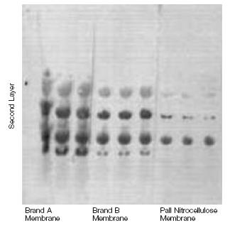
Prestained proteins were separated in a polyacrylamide gel and electrophoretically transferred to the indicated nitrocellulose membranes. A double layer of membrane was used, one directly against the gel, followed by the second layer. Signal intensity on the second layer is indicative of burn through, which can lead to loss of signal.
Multiplex Fluorescent Detection on Nitrocellulose Membrane

Human plasma spiked with myoglobin (top row) at 100, 50, 25, 10, 5, 2.5 ng per lane and Troponin I (bottom row) at 2.5, 5, 10, 25, 50 and 100 ng per lane. Plasma was diluted 10x with PBS and added to gels. After electrophoresis, proteins were electrotransferred to nitrocellulose membrane. Membranes were blocked with casein and incubated with primary antibody cocktail: donkey anti-troponin IgG and goat anti-myoglobin IgG. Following a brief wash, membranes were incubated with fluor labeled 2nd antibody: rabbit anti-donkey IgG (alexa 488 label) and rabbit anti-goat IgG (allophycocyanin label). Membranes were scanned immediately after incubation with 2nd antibody.
Type
Use
Ordering Information
Custom roll, sheet, and disc sizes available upon request. Please contact you local sales representative for additional information.
| Part Number | Description | Pkg |
| S80209 | BioTrace NT Nitrocellulose membrane, 0.2 µm, 8" x 10" sheet | 1/pkg |
Reviews
Earn 10% off* your next order online by leaving a review of this product. Please login to your account to leave a review. We appreciate and value your feedback.
*Subject to Terms and Conditions.